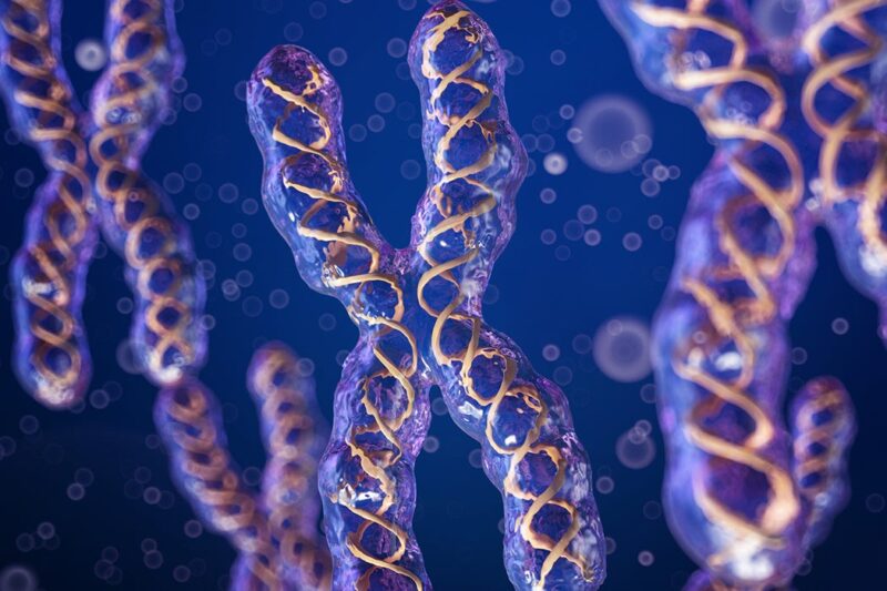
The X chromosome is one of the two sex chromosomes in humans (the other is the Y chromosome). The sex chromosomes form one of the 23 pairs of human chromosomes in each cell. It spans about 155 million base pairs (the building blocks of DNA) and represents approximately 5 percent of the total DNA in cells. Each person normally has one pair of sex chromosomes in each cell. Females have two X chromosomes, while males have one X and one Y chromosome.
Early in embryonic development in females, one of the two X chromosomes is randomly and permanently inactivated in somatic cells (cells other than egg and sperm cells). This phenomenon is called X-inactivation or Lyonization. X-inactivation ensures that females, like males, have one functional copy of the X chromosome in each body cell.
Because X-inactivation is random, in normal females the X chromosome inherited from the mother is active in some cells, and the X chromosome inherited from the father is active in other cells.
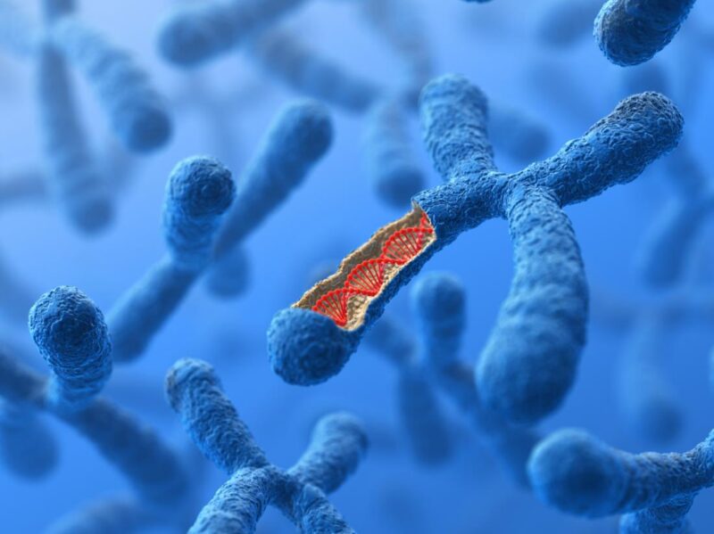
Some genes on the X chromosome escape X-inactivation. These genes are located at the tip of the short (p) arm of the X chromosome in an area known as the pseudoautosomal region. Although many genes are unique to the X or Y chromosome, genes in the pseudoautosomal region are present on both chromosomes.
As a result, men and women each have two functional copies of these genes. Many genes in the pseudoautosomal region are essential for normal development.
Identifying genes on each chromosome is an active area of genetic research. Because researchers use different approaches to predict the number of genes on each chromosome, the estimated number of genes varies. It likely contains between 900 and 1,400 genes. Genes on the X chromosome are among the estimated 20,000 to 25,000 total genes in the human genome.
What Chromosomal Conditions Are Related to The X Chromosome?
Changes in the structure or number of copies of a chromosome can also cause problems with health and development. The following chromosomal conditions are associated with such changes in the X chromosome.
Intestinal Pseudo-Obstruction
Intestinal pseudo-obstruction, a condition characterized by impairment of the muscle contractions that move food through the digestive tract (peristalsis), can be caused by genetic changes within the X chromosome.
Some individuals with intestinal pseudo-obstruction have mutations, duplications, or deletions of genetic material in the X chromosome that affect the FLNA gene. Researchers believe that these genetic changes may impair the function of the filamin A protein, causing abnormalities in the cytoskeleton of nerve cells (neurons) in the gastrointestinal tract.
These abnormalities result in impaired peristalsis, which causes abdominal pain and other gastrointestinal symptoms of intestinal pseudo-obstruction. Deletions or duplications of genetic material that affect the FLNA gene can also include adjacent genes on the X chromosome.
Changes in adjacent genes may account for some of the other signs and symptoms, such as neurological abnormalities and unusual facial features, that occur in some affected individuals.
Klinefelter syndrome
Klinefelter syndrome is caused by the presence of one or more extra copies of the X chromosome in a male’s cells. Extra genetic material from the X chromosome interferes with male sexual development, preventing the testes from functioning normally and reducing the levels of testosterone (a hormone that directs male sexual development). Typically, males with Klinefelter syndrome have one extra copy of the X chromosome in each cell, for a total of two X chromosomes and one Y chromosome (47, XXY).
Less commonly, affected males may have two or three extra X chromosomes (48, XXXY or 49, XXXXY) or extra copies of both the X and Y chromosomes (48, XXYY). In males with more than one additional chromosome, the extra genetic material may lead to mental retardation and other medical problems.
Some males with Klinefelter syndrome have the extra chromosome in only some of their cells; in these individuals, the condition is described as mosaic Klinefelter syndrome (46, XY/47, XXY).
Microphthalmia with Linear Skin Defects Syndrome
A deletion of genetic material in a region of the X chromosome called Xp22 causes microphthalmia with linear skin defects syndrome. This region includes a gene called HCCS, which carries instructions for producing an enzyme called holo cytochrome c-type synthase. This enzyme helps produce a molecule called cytochrome c. Cytochrome c is involved in a process called oxidative phosphorylation, by which mitochondria generate adenosine triphosphate (ATP), the cell’s main energy source.
It also plays a role in the self-destruction of cells (apoptosis). A deletion of genetic material that includes the HCCS gene prevents the production of the holo cytochrome c-type synthase enzyme. In females (who have two X chromosomes), some cells produce a normal amount of the enzyme and other cells produce none.
The resulting overall reduction in the amount of this enzyme leads to the signs and symptoms of microphthalmia with linear skin defects syndrome. In males, (who have only one X chromosome), a deletion that includes the HCCS gene results in a total loss of the holo cytochrome c-type synthase enzyme. A lack of this enzyme appears to be lethal very early in development, so almost no males are born with microphthalmia with linear skin defects syndrome.
A few affected individuals with male appearance but who have two chromosomes have been identified. A reduced amount of the holo cytochrome c-type synthase enzyme can damage cells by impairing their ability to generate energy.
In addition, without the holo cytochrome c-type synthase enzyme, the damaged cells may not be able to undergo apoptosis. These cells may instead die in a process called necrosis that causes inflammation and damages neighboring cells. During early development, this spreading cell damage may lead to eye and skin abnormalities characteristic of microphthalmia with linear skin defects syndrome.
Triple X Syndrome
Triple X syndrome (also called 47, XXX, or trisomy X) results from an extra copy of the X chromosome in each of a female’s cells. Females with trisomy X have three X chromosomes, for a total of 47 chromosomes per cell. It is unclear why an extra copy of the chromosome is associated with tall stature, learning problems, and other features in some girls and women. Some females with triple X syndrome have an extra X chromosome in only some of their cells.
This phenomenon is called 46, XX/47, XXX mosaicism. Females with more than one extra copy of the X chromosome (48, XXXX, or 49, XXXXX) have been identified, but these chromosomal changes are rare. The extra genetic material may lead to intellectual disability and other medical problems.
Turner Syndrome
Turner syndrome results when one normal X chromosome is present in a female’s cells and the other sex chromosome is missing or structurally altered. The missing genetic material affects development before and after birth, leading to short stature, ovarian malfunction, and the other features of Turner syndrome.
About half of the individuals with Turner syndrome have monosomy X (45, X), which means each cell in an individual’s body has only one copy of the X chromosome instead of the usual two sex chromosomes. Turner syndrome can also occur if one of the sex chromosomes is partially missing or rearranged rather than completely absent.
Some women with Turner syndrome have a chromosomal change in only some of their cells, which is known as mosaicism. Some cells have the usual two sex chromosomes (either two X chromosomes or one chromosome and one Y chromosome), and other cells have only one copy of the chromosome. Women with Turner syndrome caused by X chromosome mosaicism (45, X/46, XX or 45, X/46, XY) are said to have mosaic Turner syndrome.
Researchers have not determined which genes on the X chromosome are responsible for most of the features of Turner syndrome. They have, however, identified one gene called SHOX that is important for bone development and growth. The SHOX gene is located in the pseudoautosomal region of the chromosome. Missing one copy of this gene likely causes short stature and skeletal abnormalities in women with Turner syndrome.
Read the rest of this article from the Genetics Home Reference. – –